Synopsys and Ansys power the future of innovation—connecting silicon to systems.
-
-
Accédez au logiciel étudiant gratuit
Ansys donne les moyens à la prochaine génération d'ingénieurs
Les étudiants ont accès gratuitement à un logiciel de simulation de classe mondiale.
-
Connectez-vous avec Ansys maintenant !
Concevez votre avenir
Connectez-vous à Ansys pour découvrir comment la simulation peut alimenter votre prochaine percée.
Espace client
Support
Communautés partenaires
Contacter le service commercial
Pour les États-Unis et le Canada
S'inscrire
Essais gratuits
Produits & Services
Apprendre
À propos d'Ansys
Back
Produits & Services
Back
Apprendre
Ansys donne les moyens à la prochaine génération d'ingénieurs
Les étudiants ont accès gratuitement à un logiciel de simulation de classe mondiale.
Back
À propos d'Ansys
Concevez votre avenir
Connectez-vous à Ansys pour découvrir comment la simulation peut alimenter votre prochaine percée.
Espace client
Support
Communautés partenaires
Contacter le service commercial
Pour les États-Unis et le Canada
S'inscrire
Essais gratuits
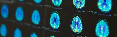
Every year, 69 million people experience a traumatic brain injury (TBI), which accounts for 39% of injury-related deaths. But these numbers only represent diagnosed cases; many mild TBIs or concussions go unreported. To help us better understand the impact of TBIs, Imperial College London is redefining our understanding of brain injuries, particularly in how mechanical forces impact brain function. The HEAD Lab, led by Dr. Mazdak Ghajari, has established itself as a leader in examining the intricate relationships between biomechanics and neurological damage, focusing on conditions like TBIs.
Using advanced computational techniques, Ghajari’s team of researchers analyze how head impacts create strain on brain tissues, often revealing insights into the onset of injuries that traditional experimental methods cannot replicate. This work extends beyond theoretical understanding; it informs the design of helmets and lays the groundwork for diagnostic tools that can be integrated into clinical settings. By leveraging high-fidelity brain models, Ghajari’s team connects the dots between real-world trauma and the resulting structural damage.
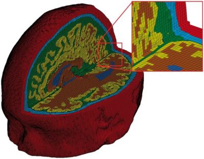
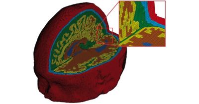
A finite element mesh of a high-fidelity 3D model of a traumatic brain injury. The colors indicate skin (red), skull (light blue), cerebrospinal fluid (green), gray matter (yellow), white matter (brown), and ventricles (dark blue). © Mazdak Ghajari (2016). Published by Oxford University Press on behalf of the Guarantors of Brain. All rights reserved.
Predictive Modeling for Patient-Specific Care
“The brain is super complicated,” says Ghajari. “No two impacts are the same. We have seen it in our simulation. When you look at the transient simulation, we can see the brain is twisting in the center. But without simulation, we are not really able to see what is inside the box. Some say we can use physical surrogates, but our computational models nowadays are far more biofidelic.”
With advanced simulation tools and detailed finite element models, researchers at the HEAD Lab analyze how individual differences in brain anatomy influence injury mechanics. Predictive modeling enables the identification of how specific brain regions are affected by mechanical forces during impacts, enabling the precise mapping of injury risk.
The team’s brain model is the result of more than a decade of development in collaboration with neurologists, enabling researchers to refine its accuracy over time and better capture the complex mechanics of brain injury.
By incorporating variables like cranial volume and sulcal depth (depth of the grooves in and around the gyri – the ridges and folds of the brain), these models reveal individual susceptibility to injury. The integration of blood biomarker analysis further enriches these predictions, providing measurable indicators that can correlate with strain patterns observed in simulations. This comprehensive perspective fosters the development of individualized care strategies, ensuring that treatments and preventive measures are more closely aligned with the unique physiological characteristics of each patient.
The adoption of Ansys simulation tools accessed through the Ansys Academic Program, particularly Ansys LS-DYNA nonlinear dynamics structural simulation software, has enabled researchers at The HEAD Lab to simulate the dynamic responses of brain tissue under impact scenarios with high precision. LS-DYNA software capabilities enable the modeling of complex, transient nonlinear phenomena, such as tissue deformation caused by traumatic forces. Using finite element analysis (FEA), researchers can evaluate how different mechanical loads translate into strain patterns within the brain, offering critical insights into the physical processes leading to traumatic brain injuries.
“LS-DYNA is extremely powerful dealing with non-linear, transient dynamics problems,” says Ghajari. “And traumatic brain injury is one of the clear examples where we have non-linearity.”
This tool's utility is amplified when integrated with machine learning techniques, which help refine simulations and enhance predictive accuracy. For example, machine learning has been instrumental in reducing computation time for these models, enabling faster iterations and improving their applicability for real-world scenarios. Ghajari’s team has further advanced the workflow by creating custom post-processing pipelines to transform simulation outputs into formats compatible with medical imaging software. This facilitates comparisons with clinical imaging techniques like MRIs, helping clinicians better interpret injury mechanisms.
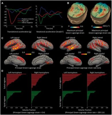
Computational results for an American football case. (A) Time history of head accelerations; (B) strain and strain rate contours within the brain; (C) the predicted strain and strain rate measured at the gray-white matter interface and overlaid onto an “inflated” brain image (gyral regions light gray and sulcal dark gray); (D) the volume fraction of sulcal and gyral regions exceeding strain and strain rate thresholds. The green bars indicate gyral regions and the red bars indicate sulcal regions © Mazdak Ghajari (2016). Published by Oxford University Press on behalf of the Guarantors of Brain. All rights reserved.
By using high-resolution anatomical data, including detailed descriptions of cerebrospinal fluid, meninges (protective membranes), and vasculature, these simulations achieve an unparalleled level of detail. The inclusion of such intricate features supports the study of tissue interactions during impacts, particularly in regions vulnerable to shear forces, such as sulci and white matter tracts. The predictive power of these simulations has been validated against clinical outcomes, demonstrating their potential to map regions of vulnerability and correlate mechanical strain with biomarkers or pathological changes. This workflow positions computational modeling as an invaluable resource in both research and healthcare applications.
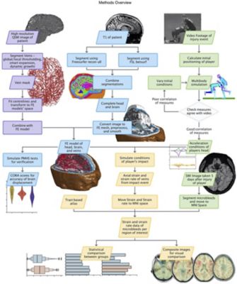
Methods flow chart with data and process boxes colored according to their theme: vein segmentation in purple, finite element model creation in blue, reconstruction in green, and results in yellow. Duckworth H, Azor A, Wischmann N, Zimmerman KA, Tanini I, Sharp DJ, and Ghajari M (2022) A Finite Element Model of Cerebral Vascular Injury for Predicting Microbleeds Location. Front. Bioeng. Biotechnol. 10:860112. doi: 10.3389/fbioe.2022.860112
Heading in the Right Direction
Researchers at Imperial College London are focusing on advancing computational models to better address the complexities of brain injury. Future efforts include enhancing real-time simulation capabilities to support immediate decision-making in clinical settings. By incorporating more refined multiphysics modeling, such as linking biomechanical forces with electrophysiological responses, researchers aim to provide deeper insights into the mechanisms underlying brain injuries. Additionally, the integration of large-scale datasets, including patient-specific anatomical and biomarker data, will enable more accurate and personalized predictions of injury outcomes. The team is also exploring ways to further reduce computational time while maintaining high model fidelity, ensuring broader accessibility for clinical applications.
Collaborations across disciplines will continue to play a vital role in refining the use of these models for injury prevention, rehabilitation, and the development of protective gear.
For Ghajari, tools like Ansys are central to enabling the kind of advanced research his team is doing.
“Tools like Ansys make it possible for us to study what’s happening inside the brain in ways we otherwise couldn’t, and we look forward to exploring their further applications in our work.”
To learn more about Ansys’ support for students, educators, and researchers, visit Ansys Academic.

Just for you. We have some additional resources you may enjoy.
“Tools like Ansys make it possible for us to study what’s happening inside the brain in ways we otherwise couldn’t, and we look forward to exploring their further applications in our work.”
— Dr. Mazdak Ghajari, associate professor in brain biomechanics, Imperial College London
The Advantage Blog
The Ansys Advantage blog, featuring contributions from Ansys and other technology experts, keeps you updated on how Ansys simulation is powering innovation that drives human advancement.